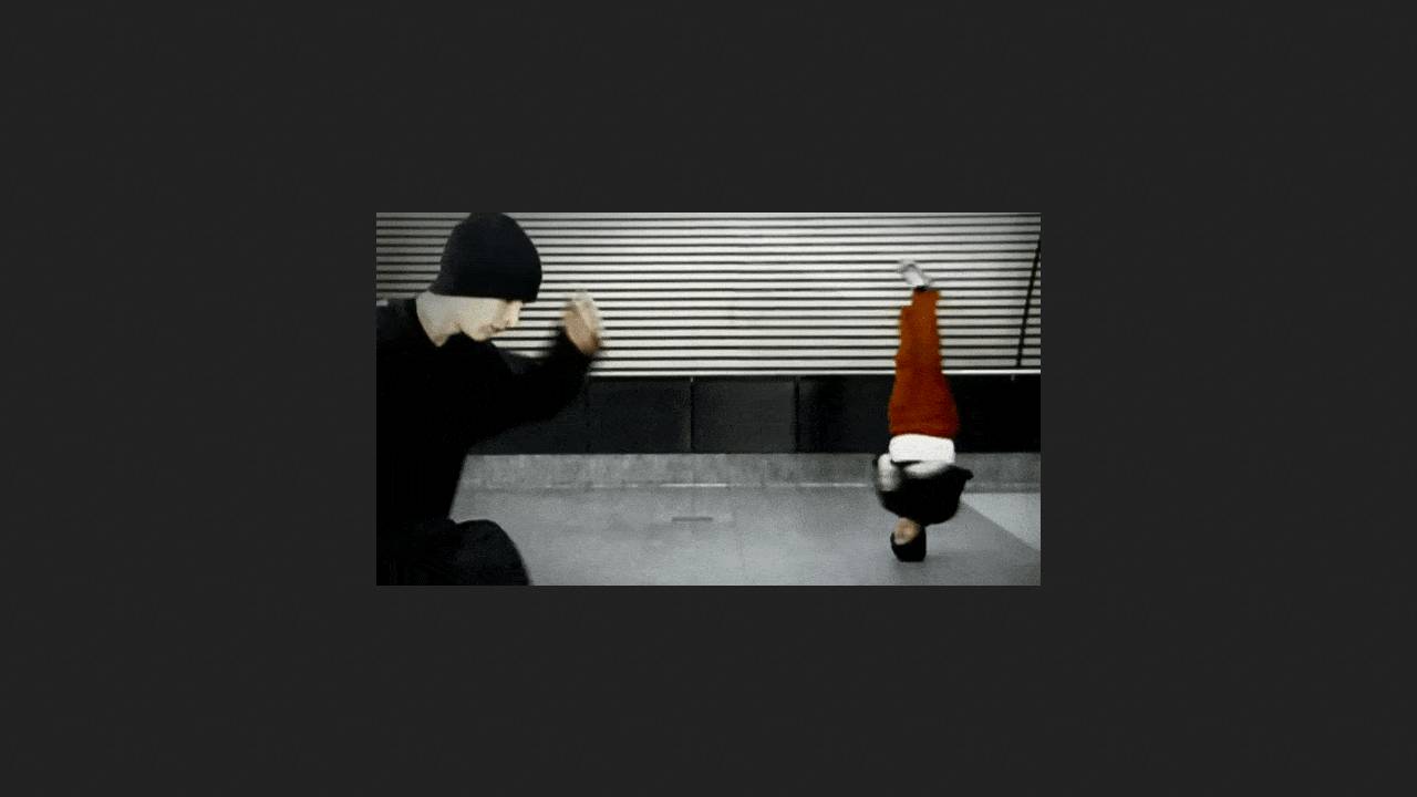
Danish doctors described a case of a painful scalp lesion causing hair loss. A medical history revealed that the patient, a professional breakdancer, had been performing headspin, a technique in which a person balances on their head while spinning around a vertical axis, for 19 years. An examination revealed that the lump was a typical case of a "headspin hole," a condition in which connective tissue grows in the scalp due to constant strain. The case is described in BMJ Case Reports.
Breakdancing involves a variety of complex and physically demanding techniques that isolate specific parts of the body. The complex nature of these movements makes breakdancers particularly susceptible to injuries to the wrists, fingers, knees, shoulders, lower back, elbows, neck, ankles, feet, and hips. The most common injuries are ligament and tendon strains.
Long-term breakdancing often leads to a number of chronic conditions, collectively known as "excessive breakdancing syndrome." This includes carpal tunnel syndrome, tendovaginitis, impingement syndrome, chronic back and head bruises, persistent hair loss, and persistent scalp irritation from headspinning. A specific form of excessive scalp damage resulting from repeated head spinning is called a "headspin hole." It is believed that up to 60 percent of dancers suffer from this condition. However, there are few case reports.
A man in his 30s was referred to doctors led by Christian Baastrup Søndergaard from the University of Copenhagen with complaints of a painful swelling of the scalp and hair loss, which had developed after 19 years of intensive breakdancing.
His regimen consisted of approximately five 90-minute workouts per week. During each workout, he performed headspins lasting between two and seven minutes. Over the past five years, the tumor had noticeably increased in size and become painful. The presence of the growth and the associated discomfort were aesthetically unpleasing to the patient, but the lump did not prevent him from continuing his breakdancing.
On clinical examination, a longitudinally oriented mass was visible on the vertex, along the midline. The mass was tender to the touch and not adherent to the skin. This led doctors to suspect that it was located beneath the caput tendineae. Magnetic resonance imaging revealed a mass beneath the caput tendineae, measuring 33.4 x 0.6 x 2.9 cm, near the midline with vascular structures. The skin and subcutaneous tissue overlying the mass were thickened, as was the underlying cranial bone.
Doctors surgically removed the tumor. Histological analysis revealed nonspecific reactive changes in the form of fairly extensive fibrosis (connective tissue proliferation) without signs of malignancy. A month later, the patient complained of no pain in the original location and was completely satisfied with the aesthetic results of the surgery.
Not only doctors but also physicists deal with dancing and its consequences. For example, our editor tried to figure out whether moshing can be described in terms of physical processes.