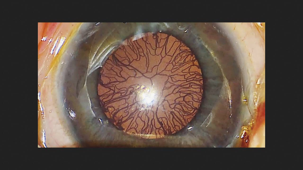
An eight-week-old infant was admitted to a Chinese ophthalmology clinic for examination and treatment. He was born at 25 weeks of gestation and weighed 850 grams at birth. Immediately after birth, he was hospitalized in the neonatal intensive care unit with respiratory distress syndrome. An ocular examination at seven weeks revealed retinopathy of prematurity. Clinical examination revealed neovascularization of the iris, as well as dilated and tortuous retinal vessels in both eyes. The blood supply extended to the posterior retina, indicating severe disease. Intraocular pressure was normal. Doctors Xinyu Zhao and Guoming Zhang from Shenzhen Eye Hospital shared this case in The New England Journal of Medicine.
Specialists decided to inject a vascular endothelial growth factor (anti-VEGF) inhibitor into the eyes to stop the growth of new vessels. Plastic dilators were inserted into the eyes, revealing a spider-like vascular network densely covering the anterior lens capsule in the anterior chambers of both eyes. Retinopathy of prematurity is characterized by slow retinal vascular development, followed by increased proliferation (presumably due to premature exposure to light and oxygen) and their invasion of areas of the eye where they normally should not be. Without timely treatment, this disease leads to blindness. An anti-VEGF injection followed by laser therapy at 17 weeks of age halted the progression of the retinopathy and stabilized the retina.