Japanese dentists described a case of a tooth forming in a child's nasal cavity. The tooth was discovered during a routine examination and was not causing any discomfort, but to prevent future inflammation, surgeons removed it through the nose under general anesthesia. The case, described in BMJ Case Reports, is rare not only because the tooth was located in the nose but also because it was not connected to the maxilla, but rather attached to the nasal septum as a polyp.
Polyodontia is a condition in which a person develops teeth beyond the normal set. It occurs in 0.1-1% of the population and is caused by a disruption in the development of tooth germs. These extra teeth are usually isolated and typically located adjacent to the primary set of teeth. Rarely, teeth are found in the nasal cavity: only 80 such cases have been described in the English-language literature, with only 14 of these teeth not having contact with bone. These extra teeth often present no obvious symptoms and are only detected by radiography. Over time, they can become inflamed or obstruct nasal breathing, so they are usually removed.
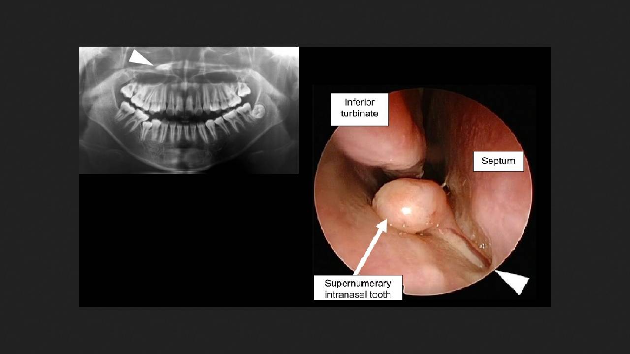
Himiko Umezawa of Saisekai Yokohamashi Nanbu Hospital and colleagues described a case of a child with an intranasal tooth. The elementary school-aged boy was brought to the dental department for malocclusion, and during a routine orthopantomography, a tooth-like lesion was discovered in his right nasal cavity. The boy did not complain and breathed freely through his nose. A visual examination by an otolaryngologist revealed no abnormalities, and a dentist confirmed the malocclusion, but all his other molars were in place.
A CT scan revealed a tooth with its crown facing the pharyngeal side of the nasal cavity base. The tooth was located in the nose without contact with the maxillary bone. Based on the CT scan, the boy was diagnosed with an additional intranasal tooth, and surgery was planned.
Surgeons removed the tooth endoscopically through the nasal cavity under general anesthesia. It was discovered that it was attached to the nasal septum as a polyp. The tooth was fully formed and reached 15 millimeters in length. The patient was monitored for a month after the surgery; the boy experienced no complications.
The exact cause of ectopic teeth is unknown. The authors believe it occurs due to excessive activation of the dental lamina, causing the tooth bud to separate from the main part of the lamina and migrate into the nasal cavity. In this clinical case, it is also interesting that the tooth was not connected to the bone in any way, but was located as a nasal polyp.
Using imaging techniques, doctors find more than just extra teeth. Sometimes, the findings are even more unusual: for example, dental implants in the maxillary sinus or a nail in the brain. Read about these and other strange CT scan findings in our article "Surprises in a Nutshell."