Physicists have created a spin qubit for quantum sensing based on a yellow fluorescent protein. The sensor's magnetic field sensitivity was 98 picoteslas at room temperature, and its coherence time was approximately 16 microseconds. The scientists implanted the resulting qubit into human embryonic kidney cells without reducing the coherence time or affecting the sensor's sensitivity. A preprint of the study is available on arXiv.org.
Qubits are not only components of quantum computers but also quantum sensors, whose operating principle is based on interaction with the environment (for example, a small change in temperature dramatically alters the coherence time and spectral characteristics of a qubit). Such sensors allow scientists to measure nanoscale electric and magnetic fields, as well as temperatures close to absolute zero, with high precision. However, the use of quantum sensors in the life sciences remains at the conceptual level today.
Most often, researchers use spin qubits based on nitrogen vacancies in diamond for biological sensing (we discussed qubits based on NV centers in more detail in the article "Quantum Technologies. Module 4"). These qubits are easily tuned optically and maintain coherence at room temperature. However, nanodiamond qubits suffer from several drawbacks critical for biological research: firstly, their large size, and secondly, their morphological inhomogeneity, which complicates their labeling.
Physicists from the United States, led by Peter Maurer of the University of Chicago, have proposed using a fluorescent protein-based qubit as a quantum sensor. To do this, the scientists used a yellow fluorescent protein (YFP) isolated from the jellyfish Aequorea victoria, in which a photoactive organic fluorophore exists in a metastable state and can be used as a spin triplet. The physicists initialized the protein's spin using optical pulses at a wavelength of 488 nanometers and used confocal microscopy to characterize the triplet state.
It turned out that in this experimental configuration, the protein remains coherent for a very short time, which is insufficient for a detailed study. Therefore, the scientists changed their approach: using an optical pulse at a wavelength of 912 nanometers, they converted the qubit from the triplet state T1 to the higher-energy triplet T2. The authors measured the coherence time of this state using the spin echo method and found that, depending on the applied magnetic field, the coherence time ranged from 140 nanoseconds to 16 microseconds. The physicists also demonstrated the potential of the developed qubit as a magnetic field sensor with an accuracy of 98 picoteslas at room temperature (for comparison, a proton five nanometers away from the qubit creates a field of 20 nanoteslas), which was made possible by the linear sensitivity of the spin contrast to the external magnetic field.
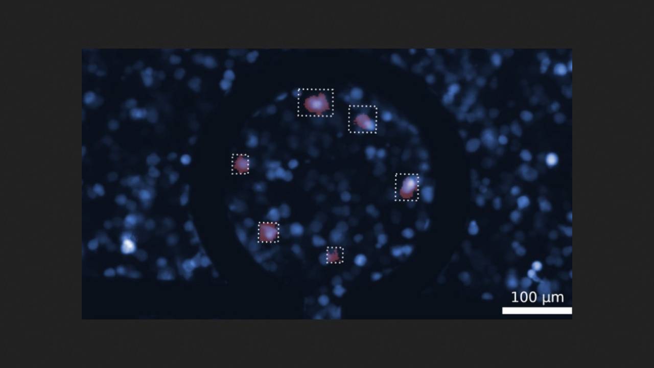
Furthermore, the scientists studied the feasibility of embedding the created qubit into mammalian cells, using human embryonic kidney cells as an example. Fluorescence imaging showed that the protein remained localized within the cells and was sensitive to an external magnetic field.
The authors of the study noted that their results have created a promising platform for quantum sensors with potential applications in biology. However, additional methods are needed to improve the qubit's coherence time and magnetic field sensitivity to achieve an advantage over existing technologies.
We previously wrote about how a diamond-based quantum sensor helped measure neural activity in the brain on a microscale.