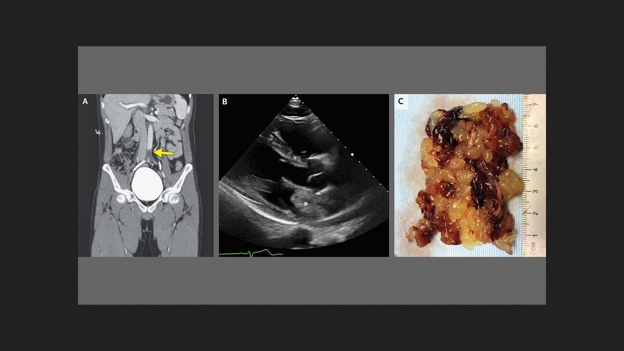
A 26-year-old man was admitted to the emergency department of an American clinic complaining of acute, severe leg pain and the inability to move his left leg. He had no previous health problems. Upon examination, he showed complete loss of motor function in his left leg, and the dorsal pedicle pulse was poorly palpable on both sides. A color Doppler ultrasound revealed a lack of blood flow in the distal aorta. A computed tomography angiography revealed a saddle embolism at the aortoiliac junction. The patient underwent an emergency embolectomy, during which a gelatinous mass was removed from the vessel. Chirag Buch and Nilam Soni, physicians at the University of Texas Health Science Center at San Antonio, shared this case in the New England Journal of Medicine.
A subsequent routine echocardiogram revealed a heterogeneous mass in the left atrium. The man underwent surgery, and a loose villous growth was removed from this atrium. Histological examination revealed abundant mucopolysaccharide matrix with scattered cellular islands. Based on these findings, the patient was diagnosed with acute abdominal aortic occlusion due to an embolism from a fragment of a left atrial myxoma—a relatively rare benign cardiac tumor. The man's recovery was uneventful, and by the time of his follow-up examination two months later, he had already returned to work.