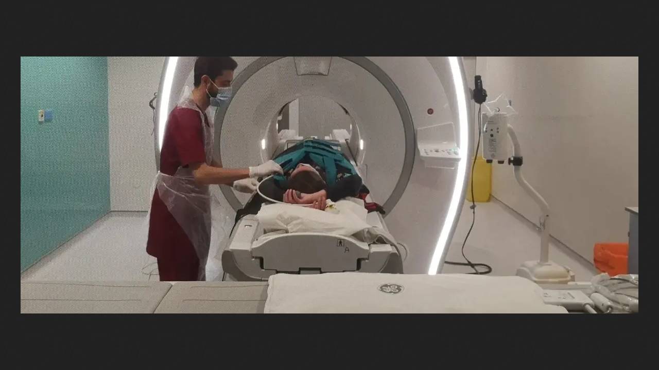
A team from Newcastle University reports a new lung imaging technology for a more accurate assessment of lung function. The examination is a two-stage process: first, patients are asked to inhale a special, harmless gas called perfluoropropane several times, followed by a standard magnetic resonance imaging (MRI) scan of the lungs.
The scientists chose perfluoropropane because it is visible on MRI scanners. This allows doctors to see which parts of the lungs are receiving oxygen and which are not.
"Signs of non-uniform ventilation are very important for early diagnosis, assessing the consequences of a progressive chronic disease, or, for example, determining lung function after a recent transplant," the scientists explained.
To date, they have confirmed the high diagnostic efficiency in patients with asthma and chronic obstructive pulmonary disease, as well as in several lung transplant recipients. The authors hope their approach will soon be widely adopted, improving the quality and speed of disease detection.
Meanwhile, the world's first fully robotic lung transplant was performed in the United States. The da Vinci Xi system performed a double lung transplant on a 57-year-old woman with COPD.