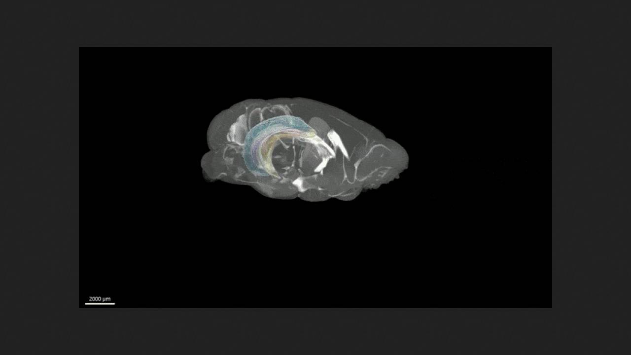
Allan Johnson of Duke University and colleagues presented a 3D stereotactic atlas of the mouse brain, covering anatomical structures and cells. To create it, the brains and skulls of five mice were imaged using three methods. First, the brains in their cranium were 3D imaged using diffusion tensor imaging at a resolution of 15 micrometers (2.4 million times higher than clinical imaging), allowing for the cytoarchitecture of brain structures to be resolved. Then, micro-computed tomography was used to mark landmarks in the skull. The brains were then removed and imaged using planar light microscopy to create cellular maps. The results were published in the journal Science Advances.
Data from all imaging methods on the brain and skull of five animals were averaged, corrected for geometric distortions, and integrated into a 3D atlas, with all structures labeled. This atlas is designed to facilitate structural and functional neurobiological studies conducted on the mouse brain and also provide visual aids for student learning. The DMBA atlas, which is approximately 13 terabytes in size, is available free of charge upon request.